Browsing Research Publications by Title
Now showing items 175-194 of 352
-
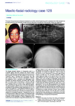
Maxillo-facial radiology case 128
(South African Dental Association, 2015)The lower clinical picture and radiographs represents a condition where the general growth is retarded, the children are pale due to anaemia and the most noticeable change of the oral structures is enlargement of the ... -
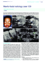
Maxillo-facial radiology case 129
(South African Dental Association, 2015)This six-year-old female patient presented with episodes of oral bleeding, fever and ulcerations affecting the submandibular region. Cropped pantomographs of the same patient were taken six weeks earlier with a follow-up ... -
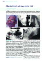
Maxillo-facial radiology case 130
(South African Dental Association, 2015)This elderly man presents with a history of a painful slow growing soft tissue swelling in his right jaw. The patient also reported paraesthesia of the right lower lip as well as paroxysms of intense general bone pain. ... -
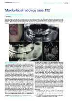
Maxillo-facial radiology case 132
(South African Dental Association, 2015)A sixteen year old female with a six year history of discomfort and pain in the left body and angle of the mandible during which time the first and second molar teeth had been extracted. Prior dental records including ... -
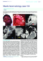
Maxillo-facial radiology case 134
(South African Dental Association, 2015)This 20 year-old male patient presented with periorbital and facial soft tissue masses of the left side of the face and a swelling affecting upper left maxilla (Figure.A, B). The patient also presents with mental retardation ... -
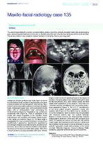
Maxillo-facial radiology case 135
(South African Dental Association, 2015)This patient presented with a sunken concave midface, anterior open bite, vertically elongated head, wide spread bulging eyes, various impacted teeth and chronic pain on the left side of the face. She also has syndactyly ... -
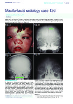
Maxillo-facial radiology case 136
(South African Dental Association, 2015)Clinical picture and skull radiographs of a patient having a developmental field defect, probably occurring between 21 and 70 days of uterine life, rather than an individual syndrome. As such the etiology and pathogenesis ... -
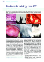
Maxillo-facial radiology case 137
(South African Dental Association, 2016)Clinical pictures and radiographs of patients presenting with a well demarcated focal mass occurring on the gingiva. Most reported series of cases show a predilection for occurrence in females with a mean age of 29 years. What ... -
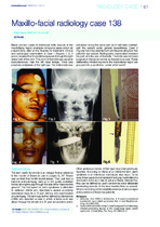
Maxillo-facial radiology case 138
(South African Dental Association, 2016)Two cases of intentional knife wounds of the maxillofacial region, examples of trauma cases which do present fairly often at the Hospital for treatment. Clinical and radiological examination of Case 1 (Figures 1, 2, 3 and ... -
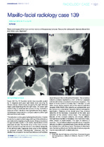
Maxillo-facial radiology case 139
(South African Dental Association, 2016)Below are images of the most common tumour of the paranasal sinuses. Discuss the radiographic features discernible and what is your diagnosis? -
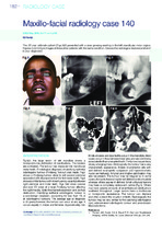
Maxillo-facial radiology case 140
(South African Dental Association, 2016)This 22 year old male patient (Figs.1&2) presented with a slow growing swelling in the left mandibular molar region. Figures 3,4,5 & 6 are images of three other patients with the same condition. Discuss the radiological ... -
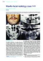
Maxillo-facial radiology case 141
(South African Dental Association, 2016)This thirty five year old male patient presented with a slow growing swelling in the right body of the mandible (Fig.1).The patient also experienced intermittent pain from time to time in the region of the swelling. What ... -
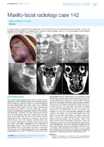
Maxillo-facial radiology case 142
(South African Dental Association, 2016)A 6-year old boy presented to the hospital with a five- month history of a persistent, draining sore (Figs.1 & 2)) in the mandible on the left side. It all started when a carious 36 was extracted. Figs. 3, 4, 5 & 6 are ... -
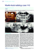
Maxillo-facial radiology case 143
(South African Dental Association, 2016)This 30 year old female presented with slight pain and discomfort and intermittent attacks of severe, itching pain in the third quadrant. The affected jaw is enlarged (Figures. 1&2). What is your diagnosis? -
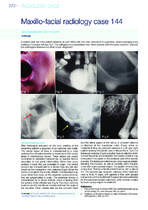
Maxillo-facial radiology case 144
(South African Dental Association, 2016)A sixteen year old male patient presents at your office with the main complaint of a painless, slowly enlarging bony swelling in his lower left jaw Fig 1. The radiographs presented are from other patients with the same ... -
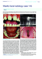
Maxillo-facial radiology case 145
(South African Dental Association, 2016)Below are clinical and radiographic images of patients who presented in the Department with the main complaint that they were not happy with the aesthetic appearance of some of their teeth which developed as they grew ... -
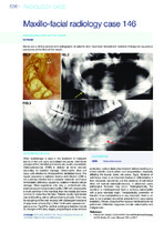
Maxillo-facial radiology case 146
(South African Dental Association, 2016)Below are a clinical picture and radiographs of patients who have been treated with radiation therapy for squamous carcinoma of the floor of the mouth. -
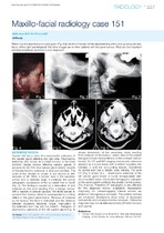
Maxillo-facial radiology case 151
(South African Dental Association, 2017)Below is a clinical picture and a radiograph (Fig.1&2) of a thirty five year old female presenting with a slow growing painless lesion of the right parotid gland. The other images are of other patients with the same tumour. ... -
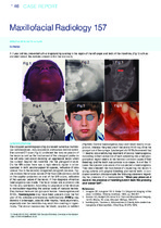
Maxillofacial radiology 157
(The South African Dental Association (SADA), 2018)The cropped pantomograph (Fig.2) showed numerous multilocular radiolucencies, bony expansion and excess tooth eruption. The coronal CT scan (Fig.3) confirmed the bony expansion of the lesion as well as the involvement ... -
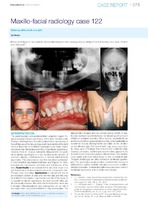
Maxillofacial radiology case 122
(South African Dental Association, 2014)Below are images of two patients with hemifacial asymmetry causing obvious disfigurement of the face and jaws. What is your diagnosis?