| dc.contributor.author | Nortje, Christoffel | |
| dc.date.accessioned | 2018-05-17T12:54:30Z | |
| dc.date.available | 2018-05-17T12:54:30Z | |
| dc.date.issued | 2016 | |
| dc.identifier.citation | Nortje, C.J. (2016). Maxillo-facial radiology case 139. South African Dental Journal, 71(3): 131 | en_US |
| dc.identifier.issn | 0011-8516 | |
| dc.identifier.uri | http://hdl.handle.net/10520/EJC188905 | |
| dc.identifier.uri | http://hdl.handle.net/10566/3690 | |
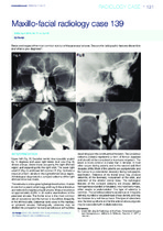
| dc.description.abstract | Below are images of the most common tumour of the paranasal sinuses. Discuss the radiographic features discernible
and what is your diagnosis? | en_US |
| dc.language.iso | en | en_US |
| dc.publisher | South African Dental Association | en_US |
| dc.rights | This file may be freely used for educational uses. No commercial reproduction or distribution of this file is permitted without written permission of the South African Dental Association (SADA). Note that the SADA retains all intellectual property rights in the article. | |
| dc.subject | Tumour | en_US |
| dc.subject | Paranasal sinuses | en_US |
| dc.subject | Osteoma | en_US |
| dc.subject | Benign bone tumour | en_US |
| dc.title | Maxillo-facial radiology case 139 | en_US |
| dc.type | Article | en_US |
| dc.privacy.showsubmitter | FALSE | |
| dc.status.ispeerreviewed | TRUE | |
| dc.description.accreditation | DHET | |