| dc.contributor.author | Nortje, Christoffelv | |
| dc.date.accessioned | 2019-10-17T11:17:13Z | |
| dc.date.available | 2019-10-17T11:17:13Z | |
| dc.date.issued | 2018 | |
| dc.identifier.citation | Nortje CJ. Maxillofacial Radiology 157. S. Afr. dent. j. [Internet]. 2018 Feb [cited 2019 Oct 17] ; 73( 1 ): 46-46. Available from: http://www.scielo.org.za/scielo.php?script=sci_arttext&pid=S0011-85162018000100010&lng=en. | en_US |
| dc.identifier.issn | 0375-1562 | |
| dc.identifier.uri | http://ref.scielo.org/j5tqq2 | |
| dc.identifier.uri | http://hdl.handle.net/10566/5044 | |
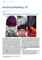
| dc.description.abstract | The cropped pantomograph (Fig.2) showed numerous multilocular
radiolucencies, bony expansion and excess tooth eruption.
The coronal CT scan (Fig.3) confirmed the bony expansion of
the lesion as well as the involvement of the pterygoid plates on
the left side (red arrow) denoting an aggressive lesion which
has spread beyond the mandible into the pterygoid muscle.
On the MRI scans there was a high intensity signal in all sequences
in both parpharyngeal fat spaces, indicative of slow
venous flow in the severely congested pterygoid plexuses. Figure
4 shows the tortuous nature of the flow-voids (arrows) visible
on the sagittal projection on one of the MRI scans, indicative
of the vascular nature of the lesion. A final diagnosis of central
haemangioma was made. The facial bones are a common site
for this very rare lesion. According to Langlais et al the literature
is inconsistent regarding the various types of vascular lesions.
They believe there are two groups of lesions: haemangioma and
AVM’s. Haemangioma may have been present since the first
year of life, whereas arteriovenous malformations (AVM’s) may
develop in a teenager, possibly after trauma. Facial asymmetry,
especially over the mandible may result from swelling or hypertrophy.
The skin or mucosa may be bluish, purplish, or reddish
(Fig.5&6). Central haemangioma may yield blood readily on aspiration,
whereas the pressure of the central AVM may drive the
plunger out of the syringe. Lamberg et al (1979) discovered that
11 deaths occurred during treatment of central haemangioma.
According to them extraction of teeth extending into a hematangiomatous
region seems to be the most common cause of fatal
bleeding, and the tooth was a molar in all cases. In 8 of the 11
cases the operator was aware of or suspected a haemangioma.
They also stressed the importance of suspecting this lesion in
young patients with gingival bleeding and mobile teeth. In conclusion
a certain clinician made the following statement regarding
the presence of a haemangioma “When you press on a
tooth in the presence of haemangioma it feels like pressing
on a soccer ball”. | en_US |
| dc.language.iso | en | en_US |
| dc.publisher | The South African Dental Association (SADA) | en_US |
| dc.subject | Multilocular radiolucencies | en_US |
| dc.subject | Pterygoid plates | en_US |
| dc.subject | Aggressive legion | en_US |
| dc.subject | Central haemangioma | en_US |
| dc.subject | Bony expansion | en_US |
| dc.title | Maxillofacial radiology 157 | en_US |
| dc.type | Article | en_US |