Browsing Faculty of Dentistry by Title
Now showing items 244-263 of 423
-
Nanobiotechnology in regenerative dental medicine
(Nanomaterials in Dental Medicine, 2023)Nanotechnology harnesses the phenomenal atomic and molecular behaviour of materials at a nanoscale size (1–100 nm) to provide solutions to a vast array of scientific applications. Although nanomaterials are commonly ... -
Neutral sodium fluoride gel uptake of newly placed nanodiamond-modified glass ionomers
(Quintessence Publishing, 2020)Three commercial restorative glass-ionomer cements (GICs) were modified with 5% and 10 wt/wt% nanodiamond (ND) particles incorporated into the powder of the GICs. The aim of the study was to assess the percentage of ... -
Neutral zone or conventional mandibular complete dentures: a randomised crossover trial comparing oral health-related quality of life
(Wiley, 2017)There is widespread consensus that the neutral zone (NZ) concept contributes to improved stability for mandibular complete dentures (CDs). However, little is known about its impact on oral health-related quality of life ... -
A Novel Dental Implant Design Concept: Radiological Bone Level Presentation of the CoAxis Dental Implant after 1 Year, And 4 Years of Prosthodontic Loading
(, 2017)Implant supported rehabilitations are routinely successful and provide patients with comfort and function levels which may surpass that of conventional treatment options. Patient-reported outcomes have revealed that ... -
Occlusal outcome of orthodontic treatment for patients with complete cleft lip and palate
(Sage Journal, 2021)Aim: To assess occlusal outcomes of orthodontic treatment for patients with complete cleft lip and palate. Design: Retrospective assessment using the Peer Assessment Rating (PAR) index. Setting: Consecutive patients ... -
Omics-based molecular techniques in oral pathology centred cancer: Prospect and challenges in Africa
(BMC, 2017): The completion of the human genome project and the accomplished milestones in the human proteome project; as well as the progress made so far in computational bioinformatics and “big data” processing have contributed ... -
Opportunities for Teledentistry in South Africa
(South African Dental Association, 2015)Information, communication and technology (ICT) is a part of everyday life, and the constant evolution of technology has resulted in many opportunities for the health care sector. While health professionals globally have ... -
Oral cancer knowledge, attitudes, and practices among dentists in Khartoum State, Sudan
(Springer Verlag, 2017)The dental professions hold an important responsibility in the control of oral cancer and the early diagnosis highly depends on their knowledge. The present study was developed to assess the knowledge, attitude, and practice ... -
Oral health and nutrition for children under five years of age: a paediatric food-based dietary guideline
(MedPharm Publications, 2013)Good nutrition is essential for good health and the development and integrity of the oral cavity. Oral health is integral to general health and essential to well-being. Dental caries is the most common oral disease in ... -
Oral health care experiences of people living with HIV in Kwazulu-Natal and Western Cape, South Africa
(Emerald Group Publishing Limited, 2015)The purpose of this paper is to ascertain the oral health experiences of people living with HIV/AIDS in the provinces of Kwazulu-Natal (KZN) and Western Cape (WC) in South Africa. Many studies have reported that people ... -
Oral health care for children attending a malnutrition clinic in South Africa
(John Wiley & Sons, Inc, 2007)Most health problems dealt with at a primary care level have an oral health impact, making it vital for oral health services to find means to integrate with other facility-based programmes at primary health care (PHC) ... -
Oral health practices and challenges facing parents of autistic children in the Western Cape (2021)
(Elsevier Ltd, 2024)Background: Periodontal status and oral hygiene practices are found to be deficient in autistic children. This is attributed to challenges in oral health practices at home and the ability to provide dental treatment in the ... -
Oral health practices and self-reported adverse effects of E-cigarette use among dental students in 11 countries: An online survey
(BMC Oral Health, 2022)Objectives E-cigarette use has become popular, particularly among the youth. Its use is associated with harmful general and oral health consequences. This survey aimed to assess self-reported oral hygiene practices, oral ... -
Oral health practices and self‑reported adverse effects of e‑cigarette use among dental students in 11 countries: An online survey
(BMC, 2022)E-cigarette use has become popular, particularly among the youth. Its use is associated with harmful general and oral health consequences. This survey aimed to assess self-reported oral hygiene practices, oral and ... -
Oral Health Status and Treatment Needs of Pregnant Women Attending Antenatal Clinics in KwaZulu-Natal, South Africa
(UWC, 2019)During pregnancy, the oral cavity is characterised by an acidic environment and an inflammatory response brought about by vomiting and changes in hormonal levels, respectively, thereby increasing the mother’s risk of ... -
Oral health-related quality of life among people living with HIV and HIV-negative adults in Kigali, Rwanda: A comparative cross-sectional study
(BioMed Central Ltd, 2024)Background Assessing health-related quality of life has become integral to people living with HIV (PLHIV) follow-up. However, there is a lack of data regarding the impact of oral health on quality of life, known as Oral ... -
Oral hygienists' self-perceived competence on completion of a blended learning course in local anaesthesia at a South African university
(South African Dental Association, 2018)The expanding scope of practice of oral hygienists require universities to offer courses that would enhance their skills. Aim and objectives: To determine the self-perceived competence of oral hygienists to deliver local ... -
Oral manifestations of syphilis: Report of four cases
(MDPI, 2022)Syphilis is an infectious disease caused by Treponema pallidum. Syphilis can present with an array of oral manifestations at different stages of disease progression. This article reports on four cases of syphilis with ... -
Oral manifestations of thrombocytopaenia
(Elsevier, 2018)The appearance in the mouth of haemorrhagic petechiae, ecchymoses or blood blisters with spontaneous bleeding is suggestive of a haemorrhagic disorder that may be caused either by functional impairment of platelets or ... -
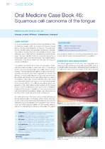
Oral medicine case book 46: squamous cell carcinoma of the tongue
(South African Dental Association (SADA), 2013)A 38-year-old-female presented at the Oral Medicine Clinic complaining of pain under her tongue that became worse during chewing, and radiated to her right ear. The pain started two months earlier and gradually increased ...